
Your shopping cart is currently empty.
Histology Prepared Microscope Slides
These prepared microscope slides can be found in the Histology Slide Kit. The microscopy images below were captured using the Richter Optica U2-D digital microscope.
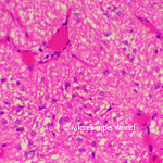 |
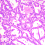
Pituitary Body The pituitary gland is an endocrine gland about the size of a pea. It is a protrusion off the base of the brain. The image of the pituitary body microscope prepared slide at left was captured at 400x magnification. Learn more about the Pituitary gland here. |
|
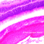 |
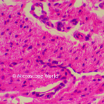
Retina Details (Choroid and Sclera) The choroid is the vascular layer of the eye containing connective tissue and lying between the retina and the sclera. The sclera is the white part of the eye and protects the outer layer of the eye. Image of retina details prepared microscope slide at left was captured at 100x magnification. You can learn more about the retina here. |
|
 |
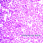
Ear (Internal Cochlea of Guinea Pig) The cochlea is the auditory portion of the inner ear. The cochlea is a spiraled, hollow, conical chamber of bone. The microscope image of the ear cochlea at left was captured at 400x magnification. You can learn more about the ear cochlea here. |
|
 |
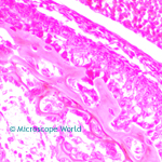
Small Intestine (with Capillaries in Villi) The small intestine is the part of the GI tract that lies between the stomach and the large intestine. The small intestine is where most of the digestion and absorption of food takes place. The prepared microscope slide of the small intestine at left was captured at 100x magnification. Learn more about the small intestine here. |
|
 |
Human Prostate Gland The prostate gland is located between the bladder and the penis. The prostate secretes fluid that nourishes and protects sperm. The prostate gland prepared microscope slide at left was captured at 100x magnification. View an anatomy picture of the prostate gland here. |
|
 |
Human Tonsil The tonsils are a pair of soft tissue masses located at the rear of the throat. The tonsils are part of the lymphatic system, which helps fight infection. The prepared microscope tonsil slide at left was captured at 400x magnification. View an image of the human anatomy of a tonsil here. |
|
 |
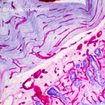
Nerve Fibers (In Bundles) Nerve fibers are threadlike extensions of a nerve cell and consist of axon and myelin sheaths in the nervous system. The nerve fiber prepared microscope slide image at left was captured at 400x magnification. |
|
 |
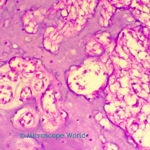
Bone and Cartilage Bones and cartilage are types of connective tissue in the body. Bones are hard tissue that form the skeletal structure in the body. Cartilages are not as hard and rigid as bones and can be found in the ear, nose and joints. In the joints, cartilage covers the ends of the bones and acts as a shock absorber to prevent the ends of bones from rubbing against each other. The bone and cartilage microscope prepared slide shown at left was captured at 400x magnification. Learn more about cartilage and bone here. |